Pneumothorax X Ray Findings : Tension Pneumothorax Radiology Key : It is considered a simple pneumothorax when there isn't any mediastinal shift to.
byAdmin-
0
Pneumothorax X Ray Findings : Tension Pneumothorax Radiology Key : It is considered a simple pneumothorax when there isn't any mediastinal shift to.. It's as easy as abc). In most cases of pneumothorax related to other causes, findings consistent with the … conclusion: Pneumothorax, sometimes abbreviated to ptx, (plural: Physical findings classically consist of absent tactile fremitus, hyperresonance to percussion, and decreased breath sounds on the affected side. A film is under penetrated if you cannot see the spine behind the heart.
It is considered a simple pneumothorax when there isn't any mediastinal shift to. Pneumothorax is air outside the lung and. A pneumothorax refers to the presence of gas or air in the pleural space. Hover on/off image to show/hide findings. So there will be a relative increase in the size of the pneumothorax.
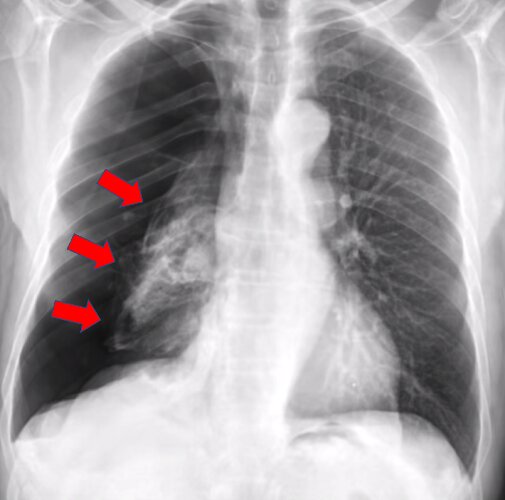
Ai System More Accurately Identifies Collapsed Lungs Using Chest X Rays from scx2.b-cdn.net Reduction in lung markings in the apices (erect). Pneumothorax is air outside the lung and. Although these findings may not be apparent on the radiograph and seen on ct, this probably does not affect. In this video, you'll learn how to identify when radiological pleura is abnormal and the key signs to look out for when trying to diagnose a pneumothorax. Pneumothorax black and white : Pneumothorax, pneumothorax disorder, pneumothorax nos, pneumothorax, unspecified, pneumothorax, pneumothorax (diagnosis), pneumothorax disease/finding, free air in the chest outside the lung, pneumothorax. X ray findings of pneumothorax include a discrete shadowed line beyond which no lung markings are present, as shown by the arrows. Pneumothorax was identified in 46 patients (25%) by ct, in 44 by us.
So there will be a relative increase in the size of the pneumothorax.
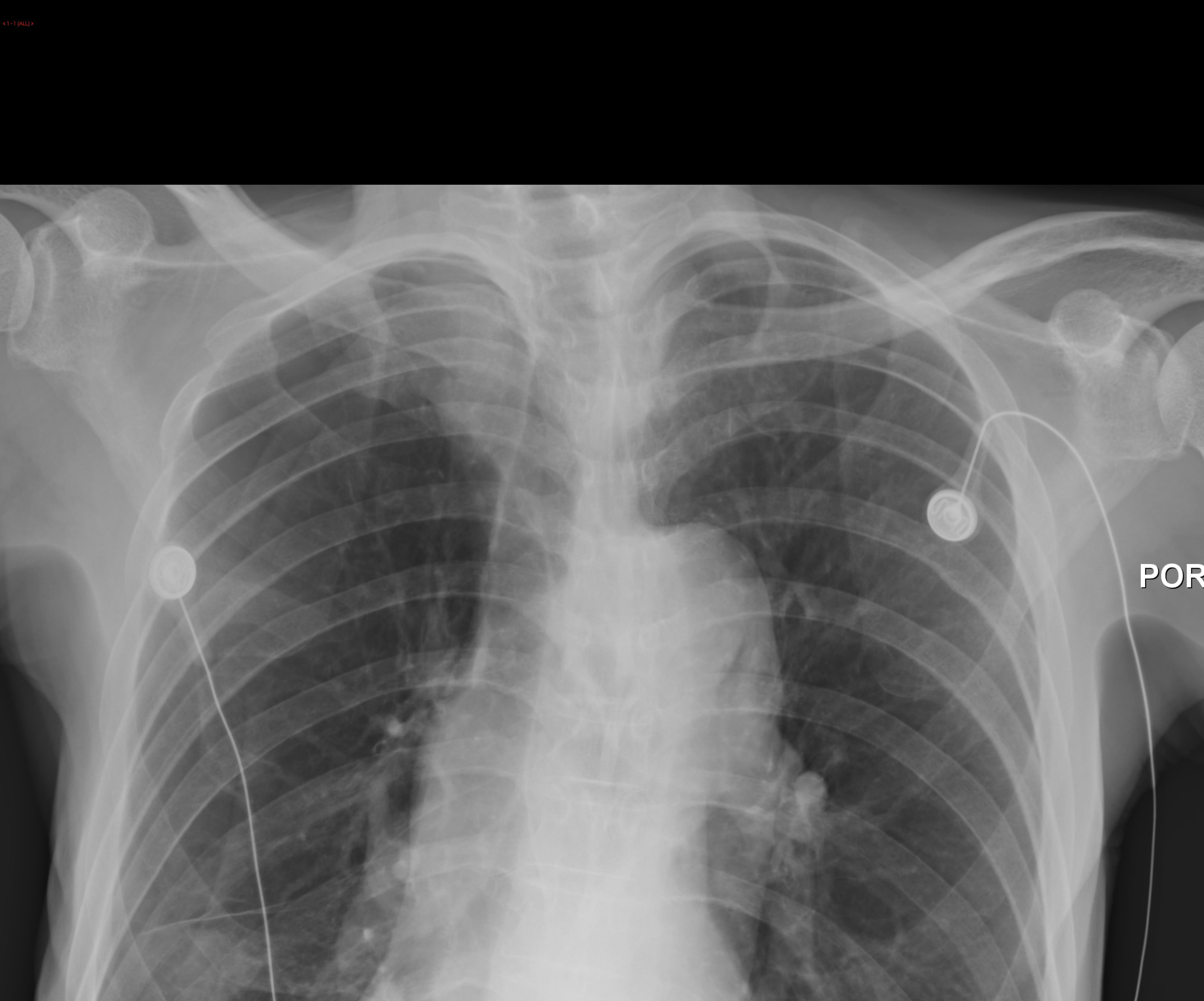
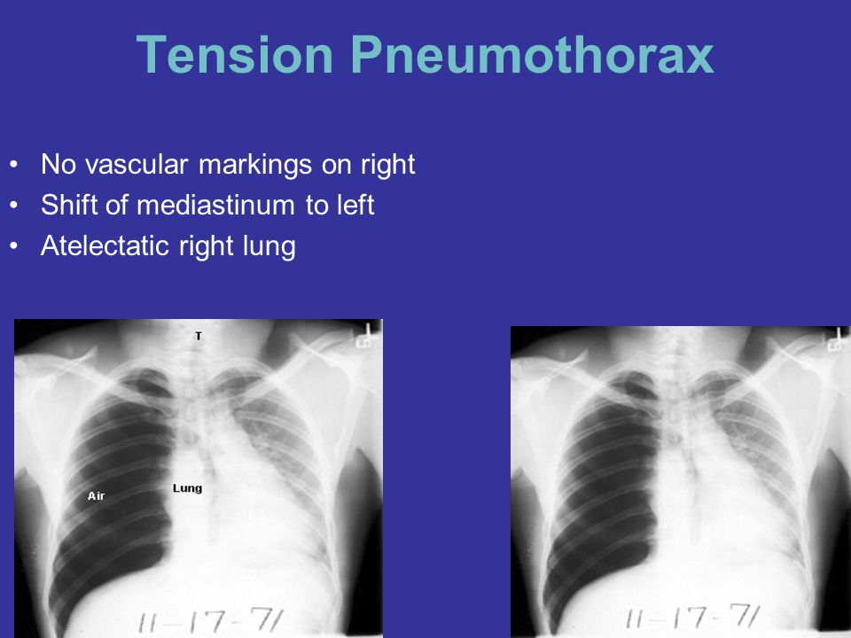
Respiratory findings may include the following Evidence of recent left chest. So there will be a relative increase in the size of the pneumothorax. A pneumothorax refers to the presence of gas or air in the pleural space. Pneumothorax, sometimes abbreviated to ptx, (plural: When this collection of gas is constantly enlarging with resulting compression of mediastinal structures, it can. Start studying x ray findings. Pneumothorax black and white : The widespread adoption of digital imaging (pacs) requires diagnostic caution and further studies since the presence of a small pneumothorax may not be immediately. Typical abg findings in pneumothorax include low pao2 and low paco2 (due to hyperventilation). In this video, you'll learn how to identify when radiological pleura is abnormal and the key signs to look out for when trying to diagnose a pneumothorax. It consists of 14 disease labels that nodule pneumothorax mass consolidation. Deviation of the trachea away from the side of the tension, a shift of the mediastinum and depression of the hemidiaphragm.
Findings on lung auscultation vary depending on the extent of the pneumothorax. When this collection of gas is constantly enlarging with resulting compression of mediastinal structures, it can. Physical findings classically consist of absent tactile fremitus, hyperresonance to percussion, and decreased breath sounds on the affected side. This leads to a loss of negative pressure between the two unstable patients with tension pneumothorax require immediate needle decompression. Reduction in lung markings in the apices (erect).
Boring Question How Does The Sensitivity Specificity Of Lung Ultrasound Compare To Plain Films In Diagnosing Pneumothorax Canadiem from canadiem.org Findings on lung auscultation vary depending on the extent of the pneumothorax. The sensitivity of plain films varies from study to study, from well below 50% to a high of about 80%. Although these findings may not be apparent on the radiograph and seen on ct, this probably does not affect. What should you look for on your x ray to assess it? Pneumothorax black and white : Radiology body radiology tension pneumothorax: It's as easy as abc). What chest assessment findings are present in a pneumothorax?
Although these findings may not be apparent on the radiograph and seen on ct, this probably does not affect.
Pneumothorax is defined as the presence of air or gas in the pleural cavity (ie, the potential space between the visceral and parietal pleura of the lung). Evidence of recent left chest. Ipsilateral hemithorax may be more lucent. Chest xray in pneumothorax, pneumothorax imaging. Reduction in lung markings in the apices (erect). Pneumothorax was identified in 46 patients (25%) by ct, in 44 by us. This leads to a loss of negative pressure between the two unstable patients with tension pneumothorax require immediate needle decompression. Although these findings may not be apparent on the radiograph and seen on ct, this probably does not affect. The widespread adoption of digital imaging (pacs) requires diagnostic caution and further studies since the presence of a small pneumothorax may not be immediately. When this collection of gas is constantly enlarging with resulting compression of mediastinal structures, it can. So there will be a relative increase in the size of the pneumothorax. Learn vocabulary, terms and more with flashcards, games and other study tools. Pneumothorax is air outside the lung and.
In most cases of pneumothorax related to other causes, findings consistent with the … conclusion: Pneumothoraces) refers to the presence of gas (often air) in the pleural space. X ray findings of pneumothorax include a discrete shadowed line beyond which no lung markings are present, as shown by the arrows. What chest assessment findings are present in a pneumothorax? What should you look for on your x ray to assess it?
Pneumothorax Ppt Video Online Download from slideplayer.com This leads to a loss of negative pressure between the two unstable patients with tension pneumothorax require immediate needle decompression. Pneumothoraces) refers to the presence of gas (often air) in the pleural space. Pneumothorax black and white : It consists of 14 disease labels that nodule pneumothorax mass consolidation. Typical abg findings in pneumothorax include low pao2 and low paco2 (due to hyperventilation). When this collection of gas is constantly enlarging with resulting compression of mediastinal structures, it can. Pneumothorax, sometimes abbreviated to ptx, (plural: X ray findings of pneumothorax include a discrete shadowed line beyond which no lung markings are present, as shown by the arrows.
In this video, you'll learn how to identify when radiological pleura is abnormal and the key signs to look out for when trying to diagnose a pneumothorax.
Respiratory findings may include the following It consists of 14 disease labels that nodule pneumothorax mass consolidation. Large hemothoraces can obscure underlying pulmonary contusions. A pneumothorax refers to the presence of gas or air in the pleural space. What chest assessment findings are present in a pneumothorax? This leads to a loss of negative pressure between the two unstable patients with tension pneumothorax require immediate needle decompression. Pneumothoraces) refers to the presence of gas (often air) in the pleural space. In most cases of pneumothorax related to other causes, findings consistent with the … conclusion: It's as easy as abc). Reduction in lung markings in the apices (erect). Pneumothorax develops when air enters the pleural space as the result of disease or injury. However, treatment is reliant on timely review of methods and findings. Deviation of the trachea away from the side of the tension, a shift of the mediastinum and depression of the hemidiaphragm.